The biology of disease can be confounding. While some single-source diseases can be accurately predicted and have a clear onset and progression path, many of the most common illnesses are incredibly complex.
Take cancer. We have multiple treatments for it, but with its dynamic tumor growth and constant evolution, we have yet to grasp what it is that can stop it.
Methods to unravel this complexity produce such vast amounts of data to truly cover all aspects of a disease – how it is initiated, molecular pathways involved in progression, our immune response, cell types involved in resolution – that every step involves its own processes, each cascading from the next, that are always changing depending on our response and the microenvironment they are part of.
This is particularly important in efforts to develop safe and effective medicines. Understanding how a disease progresses can provide potential mechanisms of action that can then be the target of a new drug. Deciphering this information using the latest technologies in cytometry empowers users to ask broader questions and leverage more quantitative analysis that can accelerate studies, moving from sample to results faster.
Immune profiling the intricacies of immune response
The immune system is integral to our health and plays a key role in therapeutic success. Yet, because of variations in immune systems across the human population, individuals can respond very differently to the same treatment. A deeper understanding of immune cell activity in relation to treatment efficacy and safety can help us predict positive response from those who get no benefit from a medicine, ultimately enabling the development of better, more targeted treatments.
How can we thoroughly and systemically delve into these processes?
Immune profiling offers a snapshot of a person’s immune health status. Applying this approach longitudinally helps us gain a more comprehensive view of disease progression and immune response. It is an invaluable technique to assess the immune system, especially to
- discover biomarkers to predict responders to therapy;
- monitor onset of disease or response to treatment;
- gain a deeper understanding of the immune system and its role in multiple disease states.
The more we can get from immune profiling about the action of a novel therapeutic, the more likely we are to be able to determine its success.
Current challenges in immune profiling
Even though technologies have advanced to enable the generation of more data, initiation of new programs and investment in updating or fully replacing protocols can be time-consuming. However, once implemented, these newer technologies can have a far greater impact and significantly improve not only the drug development process but also how and what we can learn from the underlying biology we can newly observe.
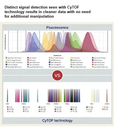
For example, conventional flow cytometry is a very low-parameter approach, with the ability to detect just 2–10 markers at a time, and the higher parameter you go, the more tedious and complex the assays become. Since the technology is based on fluorescent markers, there is a significant challenge in balancing co-expression, signal overlap and fluorophore brightness, all of which influence the number of parameters that can be measured.
Depending on the level of protein expression targeted by each antibody, the fluorophore type must be matched to a protein with an expression level such that no fluorophore will be too bright to cover other signals or so dim as to go undetected. In an experimental or clinical/translational setting, expression of key markers such as for checkpoint or signaling molecules can be quite unpredictable, adding to the complexity of panel design. These issues also necessitate additional validation and staining controls, adding more steps and tubes of sample for every experiment. As one would surmise, the more fluorophores used, the more antibody management needed and the more difficult it is to build or modify larger panels.
While several challenges must be managed when using conventional and spectral flow cytometry for immune profiling, mass cytometry empowers scientists to overcome interfering factors such as workflow standardisation, preparation and storage of cells for later analysis, autofluorescence, sample heterogeneity, data quality and post-processing requirements.
An increased need to discern cell phenotype and function simultaneously in a multiplexed fashion necessitates these types of improvements in speed and resolution of single-cell technologies. Suspension-based cytometry, refined to meet the required levels of sensitivity and reproducibility for adequate measurement, can be used to monitor the immune system in assessing efficacy of different treatments and gauge success of novel immunotherapies.
High-parameter cytometry with CyTOF technology
As a more advanced approach to immune profiling, mass cytometry, the basis of CyTOF® instruments, immediately offers a paradigm shift in what you can do and how much you can learn from one sample. The technology uses pure metal-tagged antibodies and mass spectrometry to resolve highly multiplexed protein markers (both surface and intracellular) and reveals systems-level biology at single-cell resolution.
This powerful yet simple single-detector system is built with the capability to use 135 channels and a straightforward signal path from ionisation to detection that results in negligible background and minimal signal overlap. The ability to match metals to antibodies without worrying about color combinations allows addition or removal of markers with ease. What these features translate to is the ability to quickly build larger panels and a high degree of inter-site reproducibility and intermediate precision, minimising variability from different technicians, instruments or day-to-day operations.
The use of high-parameter tools allows a look at everything at once, making the most efficient use of limited samples and providing an unbiased view of underlying biology. Whether you want to see cell type by surface markers or activation status by intracellular markers, CyTOF panels empower bigger questions to be asked and solutions realised. This equates to true high-parameter analysis, demonstrated in over 2,000 publications and more than 200 clinical trials.
Why high-parameter cytometry for drug development
The ability to ask broader questions using CyTOF technology is due to its inherently high-parameter capabilities. For example, non-targeted effects can stop a novel drug candidate from moving forward. The capability to extensively screen for non-targeted effects offers insight into efficacy data and enables further analysis, such as translational modeling and simulation, to determine if a target is direct or is being taken up by non-targeted cell populations.
More quantitative analysis facilitates receptor quantification and receptor occupancy assays to assess binding of therapeutics to their targets and advise dosing decisions, while retaining the ability to deeply phenotype complex biological systems. This helps better inform a project and generates additional information that empowers teams to expand the questions that they can ask.
Accelerating drug development with multiplexing and standardisation
Multiplexing outputs and simultaneously detecting stimulation states of cells within different immune subpopulations broaden data generation and accelerate a lab’s ability to do more and deliver therapeutics faster.
For example, cell barcoding using CyTOF technology enables multiplexing, improves workflows and enhances data quality. Barcoded samples are stained together in a single tube, eliminating staining variability between samples and reducing antibody and reagent use. Acquisition time is faster, requiring only a single run of the multiplexed sample. This allows for higher sample throughput in scaled-up experiments and improved data consistency for large studies.
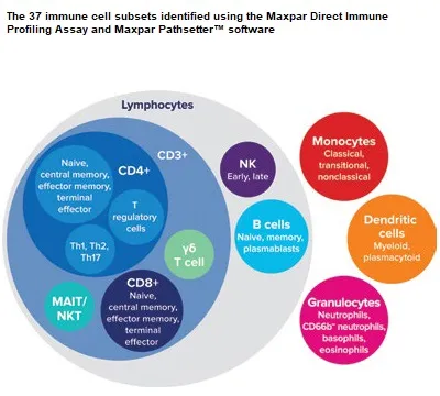
CyTOF workflows and instrumentation also allow for the standardisation of immune profiling assays with more automated sample acquisition to help reduce error and variability. They also offer the ability to prepare, stain and store samples for batch analysis at a later date or for shipping to a central facility. The fact that more than 50 markers can be confidently identified in a single experiment without having to run validation or staining controls enables a deeper dive into cell types and their functions while maximising small sample volumes. Additionally, the Maxpar® Direct™ Immune Profiling Assay™, a singletube assay measuring 37 cell populations from a 30-marker antibody panel, improves standardisation of protocols since sample (human whole blood or PBMC) is added directly to the tube and no assayspecific optimisation is required.
CyTOF technology provides impressively high resolution of cytometric profiles, empowering basic research and practical biomarker-driven clinical/translational research to optimise and accelerate drug development. CyTOF instruments are proven valuable additions to the drug development pipeline, aiding in the creation of a framework to integrate knowledge and build a foundation on which drug development and treatment strategies can rely, ultimately leading to effective medicines and potential cures.